What is the principle of immunoprecipitation?
The principle of immunoprecipitation (IP) is based on the specific binding of an antibody to its target antigen. Antibodies are proteins produced by the immune system that specifically recognize and bind to specific antigens, such as proteins or peptides. In IP, the antibody is used to specifically pull out the target protein of interest from a mixture of proteins.
The process of IP involves adding an antibody that specifically binds to the target protein to a cell lysate or tissue extract, which contains the mixture of proteins. The mixture is then incubated to allow the antibody to bind to the target protein. The mixture is then passed over a column containing a solid support, such as agarose beads, which is then washed to remove unbound proteins. The target protein is eluted from the column and can be analyzed by various methods, such as Western blotting, mass spectrometry, or enzymatic assays.
The specificity of IP is crucial, as the antibody must specifically bind to the target protein and not to other proteins in the mixture. This can sometimes be a challenge, as many antibodies can have cross-reactivity with other proteins. To ensure specificity, it is important to use high-quality antibodies and carefully validate the specificity of the IP.
What is the difference between Western blot and immunoprecipitation?
Western blot and immunoprecipitation (IP) are both techniques used in molecular biology and biochemistry to study proteins, but they have different purposes and different ways of analyzing proteins.
Western blotting (WB) is a technique used to detect specific proteins in a mixture, such as a cell lysate or tissue extract. The basic process of WB involves separating proteins by size using electrophoresis, transferring the separated proteins onto a membrane, and then using an antibody specific to the target protein to detect its presence. WB is useful for confirming the presence of a specific protein in a mixture and for determining its molecular weight.
Immunoprecipitation (IP), on the other hand, is a technique used to isolate specific proteins from a mixture. The process of IP involves adding an antibody that specifically binds to the target protein to a cell lysate or tissue extract, which contains the mixture of proteins. The mixture is then incubated to allow the antibody to bind to the target protein, and the mixture is then passed over a column containing a solid support, such as agarose beads. The target protein is then eluted from the column and can be further analyzed by methods such as Western blotting, mass spectrometry, or enzymatic assays.
In summary, Western blotting is used to detect specific proteins in a mixture, while immunoprecipitation is used to isolate specific proteins from a mixture for further analysis. Both techniques are important tools in molecular biology and biochemistry, and they are often used in combination to study the presence and properties of specific proteins in complex mixtures.
What are the steps of immunoprecipitation?
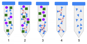
The steps of immunoprecipitation (IP) are as follows:
- Prepare cell lysate or tissue extract: The first step is to prepare a cell lysate or tissue extract, which contains the mixture of proteins to be analyzed. The lysate or extract can be prepared by breaking open cells or tissues, usually with the use of a lysis buffer, to release the proteins into solution.
- Add antibody: The second step is to add an antibody that specifically binds to the target protein to the cell lysate or tissue extract. The antibody is usually added to the lysate or extract in a buffer that contains a high salt concentration to minimize non-specific binding.
- Incubate: The third step is to incubate the mixture to allow the antibody to bind to the target protein. This is typically done at 4°C overnight or for several hours at room temperature, depending on the specificity and affinity of the antibody.
- Add solid support: The fourth step is to add a solid support, such as agarose beads, to the mixture. The solid support will be used to pull down the target protein bound to the antibody.
- Wash: The fifth step is to wash the mixture to remove unbound proteins. This is typically done by adding a high salt buffer to the mixture and then centrifuging it to remove the unbound proteins. The washing step is usually repeated several times to remove as much non-specific binding as possible.
- Elute: The sixth step is to elute the target protein from the solid support. This is typically done by adding a buffer that contains a lower salt concentration or by changing the pH of the buffer to disrupt the interaction between the antibody and the target protein. The eluted protein can then be analyzed by methods such as Western blotting, mass spectrometry, or enzymatic assays.
It is important to note that IP results must be validated to ensure specificity, as non-specific binding of the antibody or cross-reactivity with other proteins can lead to misleading results.
Contact:
David Correa
USA/Canada (Toll Free): +1-800-792-5285, +1-503-894-6022
help@alliedmarketresearch.com